
Advanced Molecular Imaging
What is
Advanced Molecular Imaging?
AMI is a medical specialty that uses radioactive tracers (radiopharmaceuticals) to assess bodily functions and to diagnose and treat disease. Specially designed cameras allow doctors to track the path of these radioactive tracers. Single Photon Emission Computed Tomography or SPECT and Positron Emission Tomography or PET scans are the two most common imaging modalities in AMI.

Radioactive tracers are made up of carrier molecules that are bonded tightly to a radioactive atom. These carrier molecules vary greatly depending on the purpose of the scan. Some tracers employ molecules that interact with a specific protein or sugar in the body and can even employ the patient’s own cells. For example, in cases where doctors need to know the exact source of intestinal bleeding, they may radiolabel (add radioactive atoms) to a sample of red blood cells taken from the patient. They then reinject the blood and use a SPECT scan to follow the path of the blood in the patient. Any accumulation of radioactivity in the intestines informs doctors of where the problem lies.
For most diagnostic studies in AMI, the radioactive tracer is administered to a patient by intravenous injection. However a radioactive tracer may also be administered by inhalation, by oral ingestion, or by direct injection into an organ. The mode of tracer administration will depend on the disease process that is to be studied. Approved tracers are called radiopharmaceuticals since they must meet FDA’s exacting standards for safety and appropriate performance for the approved clinical use. The AMI physician will select the tracer that will provide the most specific and reliable information for a patient’s particular problem. The tracer that is used determines whether the patient receives a SPECT or PET scan.
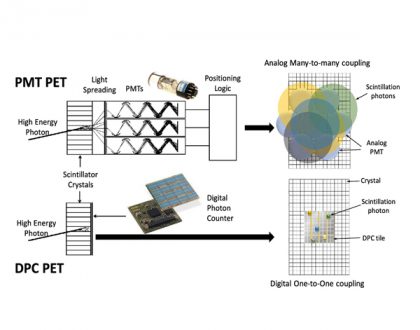
What is Single Photon Emission Computed Tomography (SPECT)?
SPECT imaging instruments provide three-dimensional (tomographic) images of the distribution of radioactive tracer molecules that have been introduced into the patient’s body.
The 3D images are computer generated from a large number of projection images of the body recorded at different angles. SPECT imagers have gamma camera detectors that can detect the gamma ray emissions from the tracers that have been injected into the patient. Gamma rays are a form of light that moves at a different wavelength than visible light. The cameras are mounted on a rotating gantry that allows the detectors to be moved in a tight circle around a patient who is lying motionless on a pallet.
What is Positron Emission Tomography (PET)?
PET scans also use radiopharmaceuticals to create three-dimensional images. The main difference between SPECT and PET scans is the type of radiotracers used.  While SPECT scans measure gamma rays, the decay of the radiotracers used with PET scans produce small particles called positrons. A positron is a particle with roughly the same mass as an electron but oppositely charged. These react with electrons in the body and when these two particles combine they annihilate each other. This annihilation produces a small amount of energy in the form of two photons that shoot off in opposite directions. The detectors in the PET scanner measure these photons and use this information to create images of internal organs.
SPECT scans are primarily used to diagnose and track the progression of heart disease, such as blocked coronary arteries. There are also radiotracers to detect disorders in bone, gall bladder disease and intestinal bleeding. SPECT agents have recently become available for aiding in the diagnosis of Parkinson’s disease in the brain, and distinguishing this malady from other anatomically-related movement disorders and dementias.
The major purpose of PET scans is to detect cancer and monitor its progression, response to treatment, and to detect metastases. Glucose utilization depends on the intensity of cellular and tissue activity so it is greatly increased in rapidly dividing cancer cells. In fact, the degree of aggressiveness for most cancers is roughly paralleled by their rate of glucose utilization. In the last 15 years, slightly modified radiolabeled glucose molecules (F-18 labeled deoxyglucose or FDG) have been shown to be the best available tracer for detecting cancer and its metastatic spread in the body.
A combination instrument that produces both PET and CT scans of the same body regions in one examination (PET/CT scanner) has become the primary imaging tool for the staging of most cancers worldwide.
Recently, a PET probe was approved by the FDA to aid in the accurate diagnosis of Alzheimer’s disease, which previously could be diagnosed with accuracy only after a patient’s death. In the absence of this PET imaging test, Alzheimer’s disease can be difficult to distinguish from vascular dementia or other forms of dementia that affect older people.

www.nibib.nih.gov
Recommended Posts
Nuclear Imaging Moves Toward Digital Detector Technology
December 10, 2019